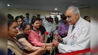










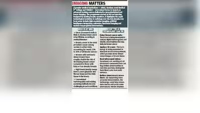
Chandigarh: Doctors at PGI have signed a memorandum of understanding with IIT Delhi to develop an indigenous artificial intelligence (AI) tool designed to detect breast cancer early in Indian women. Unlike international diagnostic tools trained on Western populations, this AI system is specifically calibrated using health data from Indian patients.“Breast cancer in Indian women often develops at a younger age — frequently appearing in their 20s and 30s — and is typically characterised by denser breast tissue, making standard mammography less effective,” said Prof Veenu Singla of the department of radiodiagnosis and imaging, PGI, who has been working on such initiatives.The project, which is currently in the trial phase, utilises a small, desktop-connected hardware box to deliver digital mammography analysis directly to a computer screen. The compact device looks like a 5 x 5-inch Rubik’s cube but is built to be roughly 1.5 times the size of a standard cube.To ensure a high degree of accuracy in the software, PGI is utilising much of its own patient data, which covers all types of breast densities, to build a robust final product. “Once fully developed and validated, the creators plan to deploy this ‘Make in India’ solution to healthcare centres across the country to improve nationwide breast cancer screening,” said Prof Veenu.The department is also working on expanding the capabilities of this technology to interpret 3D breast scans (tomosynthesis) and ultrasound videos. For the ultrasound component, the computer software will work by flagging hidden tissue abnormalities in real time, accurately separating benign fluid cysts from dangerous tumours and seeing through dense breast tissue, where traditional X-ray mammograms often fail.By providing an expert second opinion during check-ups, this technology can prevent stressful false alarms and unnecessary needle biopsies while ensuring consistent, high-standard care for every patient.Imaging MattersPremier medical institutions — AIIMS, Maulana Azad Medical College and PGIMER — have joined forces to launch an advanced training course dedicated to musculoskeletal and breast imaging. The programme started at PGI on Saturday. Designed by leading faculty members, it highlights the rapid technological evolution in radiology over the last decade. Key focal areas include high-resolution imaging, artificial intelligence integration, radiomics, functional imaging and modern image-guided interventions.Women At Risk–One in 28 women in India is likely to develop breast cancer in her lifetime, according to medical literature–Breast cancer is the most prevalent cancer among women in urban India, accounting for nearly 23% to 30% of all female cancers–Women with extremely dense breasts have roughly double the risk of developing breast cancer compared to those with fatty or low-density breasts–High breast density means there is more glandular and fibrous tissue and less fatty tissue in the breast–Conventional mammography and existing screening methods become challenging in such conditionsWhat the AI technology can do–Detect breast cancer early: The AI tool is being developed to analyse digital mammograms and identify abnormalities that may indicate breast cancer–Analyse 3D scans: The technology is being expanded to interpret breast tomosynthesis, which provides three-dimensional images of breast tissue–Read ultrasound videos: Developers are working on an ultrasound component that can flag hidden abnormalities in real time and help distinguish fluid-filled cysts from solid tumours–Reduce unnecessary procedures: By supporting more accurate interpretation, the technology could help reduce false alarms and avoid some unnecessary needle biopsies.